


 Home
Home Program
 Exhibitors
 My Meeting
 Digital Posters
 Case of Day


MRI Helps Predict Motor Deterioration in Parkinson's Disease Patients
Wednesday, Nov. 29, 2017
MRI-based measurements of the brain's white matter can help predict which Parkinson's disease (PD) patients will go on to develop motor deterioration, according to a study presented Tuesday.

Minett
Parkinson's disease is a progressive disorder of the central nervous system characterized by motor symptoms such as tremors, trembling, stiffness in the limbs and impaired balance and coordination. It affects one to two of every 1,000 people, according to the National Institutes of Health, and prevalence is rising as the population ages.
Thais Minett, MBBS, PhD, FRCR, a fourth-year trainee in Cambridge, United Kingdom, and colleagues explored the use of diffusion tensor imaging (DTI) with MRI to detect changes to the brain's white matter on a microstructural level and see if these changes are predictors of declining motor impairment in PD.
The researchers compared 120 patients with early PD and 48 healthy controls. Participants underwent clinical, motor and DTI investigations at baseline and 18 months later. The researchers analyzed DTI-generated measures of mean diffusivity and fractional anisotropy and investigated their relationships with motor function. Mean diffusivity reveals how restricted diffusion of water is within the white matter, while fractional anisotropy represents the degree of directional diffusion.
At baseline, patients with PD had significantly higher widespread mean diffusivity than controls. Baseline mean diffusivity was a significant predictor of worsening motor impairment in PD, while fractional anistropy was not a significant predictor.
"It may be that fractional anisotropy reduction only occurs later in the course of disease progression," Dr. Minett said. "Our cohort comprised of newly-diagnosed Parkinson's disease cases who had a mean disease duration of six months."
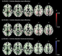
Regions at baseline of increased mean diffusivity and reduced fractional anisotropy (p <0.05 corrected). First row: in red, voxels with significantly reduced fractional anisotropy between patients with PD and controls. Bottom row: in blue, voxels with significantly increased mean diffusivity between patients with PD and controls. Results are shown overlaid on an MNI152 template and the mean FA skeleton (green). Z-coordinates are displayed. Click or tap image to enlarge.
Another possibility, she said, is that mean diffusivity measurements are inherently more sensitive than fractional anisotropy for detecting differences in white matter microstructure, as mean diffusivity is more uniform across the brain than fractional anisotropy and therefore less affected by registration errors in relation to voxel size.
"If mean diffusivity is sensitive but directionally non-specific, any process leading to white matter microstructural degeneration would cause mean diffusivity to invariably increase, whereas fractional anisotropy provides more specific information as it depends on the underlying arrangement of the fibers," Dr. Minett said.
The results suggest that mean diffusivity measurements could play a role in determining optimal care for patients with early PD, according to Dr. Minett.
"We could infer from my results that the groups of patients with higher mean diffusivity would have more motor deterioration than those with lower mean diffusivity," she said. "So, identifying a group with high likelihood of poor outcome may have important clinical relevance in terms of predicting prognosis and, therefore, the care pathway needed," Dr. Minett said. "More importantly, it may help in identifying a group with particular high need for therapeutic intervention who can be the subject of future trials."