


 Home
Home Program
 Exhibitors
 My Meeting
 Digital Posters
 Case of Day


3‑D MRI Fetal Images Superior in Quality to 3‑D US
Monday, Nov. 27, 2017
When there is a question of findings during 3-D ultrasound (US) in the third trimester of pregnancy, many radiologists are turning to 3-D MRI for anatomy exploration of the fetus and to support diagnostic decisions.
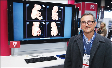
Werner
While not routinely used during prenatal care, 3-D MRI does offer excellent tissue contrast as a second evaluation in difficult cases or to reinforce an US diagnosis, according to Heron Werner, MD, PhD, at Clínica de Diagnóstico por Imagem in Rio de Janeiro.
US scanning over the past several decades has opened a new window into the study of the fetus, because it is patient-friendly, cost-effective and safe. MRI for fetal imaging has been in use since the 1980s and offers high-resolution images with excellent tissue contrast. It provides additional information about fetal abnormalities and conditions in situations where US cannot provide high-quality images, such as advanced gestational age, reduction of the amniotic fluid and maternal obesity.
"Ultrasound examination is the primary method of fetal assessment, while MRI is complementary in that it is a diagnostic technique that can provide sharp images of the human body," Dr. Werner said. "The large field of view from MRI offers the 3-D reconstruction of the whole fetal body, allowing radiologists to identify some phenotypes of different syndromes." These syndromes can include craniosynostosis, cleft lip, limb reduction, Beckwith-Wiedemann syndrome, conjoined twins and club feet.
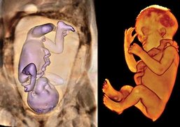
3-D MRI showing a 27-week fetus with brachycephaly, low ear implantation, syndromic profile and vestigial tail.
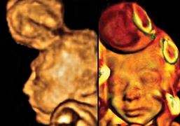
3-D ultrasound and 3-D MRI of a 23-week fetus with encephalocele.
From September 2009 to December 2016, 52 fetuses were selected from cases evaluated for external malformations. Morphological abnormalities were first imaged by 3-D US, with 3-D MRI reinforcing the preliminary findings. 3-D US scans were performed transabdominally using high-resolution US probes with harmonic images, while the MRI was a 1.5-T scanner with body coil.
The 3-D images were post-processed. Maximum intensity projection images were reconstructed and the gestational sac was manually segmented. The images were then volume rendered and the amniotic fluid was removed by threshold techniques.
Despite recent improvements in 3-D US, the results obtained from 3-D MRI were superior in the third trimester, even with fetal movements being one of the principal difficulties in capturing the images.
"For rare genetic conditions, complex malformations or even in the case of twins or triplets, 3-D MRI helps physicians understand fetal anatomical characteristics," Dr. Werner said. "3-D MRI also assists during multidisciplinary discussions among physicians who may initially differ on diagnosis or the urgency of the condition."
Ultimately, Dr. Werner foresees that 3-D MRI will be most beneficial to parents in helping them visualize their unborn baby and the challenges that the baby may face after it's born. "It is one thing to tell a parent that their baby has a tumor or serious abnormality, but it is another thing to show them," Dr. Werner summarized. "3-D MRI can help both physician and parents understand the prognosis of fetal abnormalities and help facilitate treatment decisions."